Nervous System
The nervous system includes the brain, spinal cord, and a network of nerves that extend throughout the body. The brain receives and processes information about both the external environment and internal body conditions.
The activities of the nervous system can be grouped by function:
- Sensory: The nervous system receives information from the internal and external environments through sensory receptors and processes it in the brain and spinal cord.
- Integrative: The central nervous system synthesizes and interprets information, making decisions and initiating appropriate responses. Some processing of information does occur in the peripheral nerves before entering the spinal cord.
- Motor: The brain and spinal cord send out commands that travel through efferent (motor) nerves to muscles, causing them to contract or relax, and to glands, triggering the release of various chemicals into the bloodstream.
Organization
The nervous system can be divided into the central nervous system (CNS) and the peripheral nervous system (PNS).
Central Nervous System
The central nervous system processes information from various sources and responds to internal and external stimuli. It consists of the brain and spinal cord.
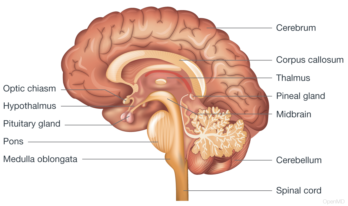
- Brain: The brain is the body’s control center. It controls a myriad of body functions, from thoughts, reasoning, and language, to regulation of breathing and heart rate. It contains close to 100 billion neurons. The brain can be divided into four anatomical areas: the cerebrum, the diencephalon, the brainstem, and the cerebellum. Each region can be further divided into specific regions and structures that each serve a unique purpose.
- Cerebrum: The cerebrum is the largest part of the brain, responsible for higher-level functions such as thought, language, cognition, emotion, creativity, and voluntary movement. It is divided into two sections called the left and right hemispheres, each of which contains five lobes, as follows:
- Frontal Lobe: The frontal lobe controls decision making, voluntary movement, attention, motivation, speech, and short-term memory tasks. It is located at the front of the brain, above and in front of the temporal lobe.
- Parietal Lobe: The parietal lobe integrates sensory input from the external world, especially touch, taste, and spatial awareness. It is situated behind the frontal lobe and above the occipital lobe.
- Occipital Lobe: The occipital lobe receives visual information and processes it. It is located at the back of the head, below the parietal lobe and behind the temporal lobe.
- Temporal Lobe: The temporal lobe processes auditory input and encodes memories. It is located at the sides of the head, below and behind the frontal lobe.
- Insula: The insula plays an important role in self-identity and various functions related to emotion or bodily regulation. It lies hidden within the lateral sulcus (a groove in the brain that separates the frontal and temporal lobes), deep to the temporal lobe.
- Cerebrum: The cerebrum is the largest part of the brain, responsible for higher-level functions such as thought, language, cognition, emotion, creativity, and voluntary movement. It is divided into two sections called the left and right hemispheres, each of which contains five lobes, as follows:
Both hemispheres are covered by an outer layer (1.5 to 4.5 mm thick) called the cerebral cortex. The left hemisphere specializes in sequential processing, language, and analytical reasoning, while the right hemisphere excels in spatial reasoning, emotional processing, and creativity. While these are general tendencies, both hemispheres interact extensively for various cognitive functions.
- Cerebellum: The cerebellum is primarily responsible for fine motor control, such as posture, balance, and coordination. It processes sensory information from the body to maintain equilibrium. Emerging research suggests that the cerebellum also contributes to certain aspects of higher cognitive functions, such as language processing and decision making.
- Diencephalon: This region of the brain includes the thalamus, hypothalamus, epithalamus, and subthalamus.
- Thalamus: Positioned between the cerebral hemispheres and the brainstem, the thalamus serves as a central relay center for sensory and motor signals. In addition, it is involved in regulating wakefulness, sleep, and awareness, and it also contributes to cognition and memory.
- Hypothalamus: Located below the thalamus, the hypothalamus is crucial for maintaining the body’s homeostasis (the body’s ability to maintain stable internal conditions for optimal functioning). It regulates temperature, hunger, thirst, and sleep cycles, and it controls hormone secretion by the pituitary gland.
- Epithalamus: This tiny brain region houses the pineal gland, which is crucial for regulating circadian rhythms (the body’s natural, internal clocks that regulate various biological processes in a roughly 24-hour cycle) and sleep patterns. Its production of melatonin varies with light exposure, influencing sleep cycles and seasonal changes.
- Subthalamus (subthalamic nucleus): From fine tuning muscle movements to coordinating complex actions, the subthalamic nucleus, a core component of the basal ganglia circuit, plays a vital role in integrating somatic motor function (the ability of the body to perform voluntary muscle movements).
- Brainstem: Consisting of the medulla oblongata, pons, and midbrain, the brainstem connects the brain to the spinal cord.
- Medulla oblongata: The medulla oblongata controls vital autonomic functions such as heart rate, blood pressure, and breathing. It also plays a role in reflex actions, such as coughing and swallowing.
- Pons: The pons is a brainstem region that links the cerebrum to the cerebellum and the medulla oblongata. It is involved in controlling sleep, arousal, focus, and awareness, as well as pain management.
- Midbrain: The midbrain, a conductor of both conscious and unconscious acts, orchestrates eye movements and auditory processing, and even plays a pivotal role in motor coordination.
- Spinal cord: A cylindrical structure composed of nervous tissue that extends from the brainstem to the lumbar region of the spinal column. The spinal cord serves as the major pathway connecting the brain to the peripheral nervous system.
Peripheral Nervous System
The peripheral nervous system consists of nerves located outside of the brain and spinal cord. The peripheral nervous system has two parts:
- Somatic nervous system: The somatic nervous system sends signals to and from the central nervous system to the skeletal muscles to mediate voluntary
- Autonomic nervous system: The autonomic nervous system controls the involuntary functions of the heart, smooth muscles, and glands. The endocrine system works with the autonomic and central nervous systems to enhance and maintain these responses, as well as other processes, such as metabolism, reproduction, and emotion. It can be further divided into sympathetic, parasympathetic, and enteric divisions.
- Sympathetic nervous system: Often referred to as the “fight or flight” system, the sympathetic nervous system prepares the body for stressful or emergency situations by increasing heart rate, dilating airways, and other physiological changes.
- Parasympathetic nervous system: Commonly associated with “rest and digest” activities, the parasympathetic nervous system conserves and restores energy by slowing the heart rate and increasing intestinal and gland activity.
- Enteric nervous system: The enteric nervous system controls gastrointestinal functions, operating independently of the sympathetic and parasympathetic systems to regulate digestive processes.
Cell Structure
The nervous system contains two distinct cell types: neurons and glial cells.
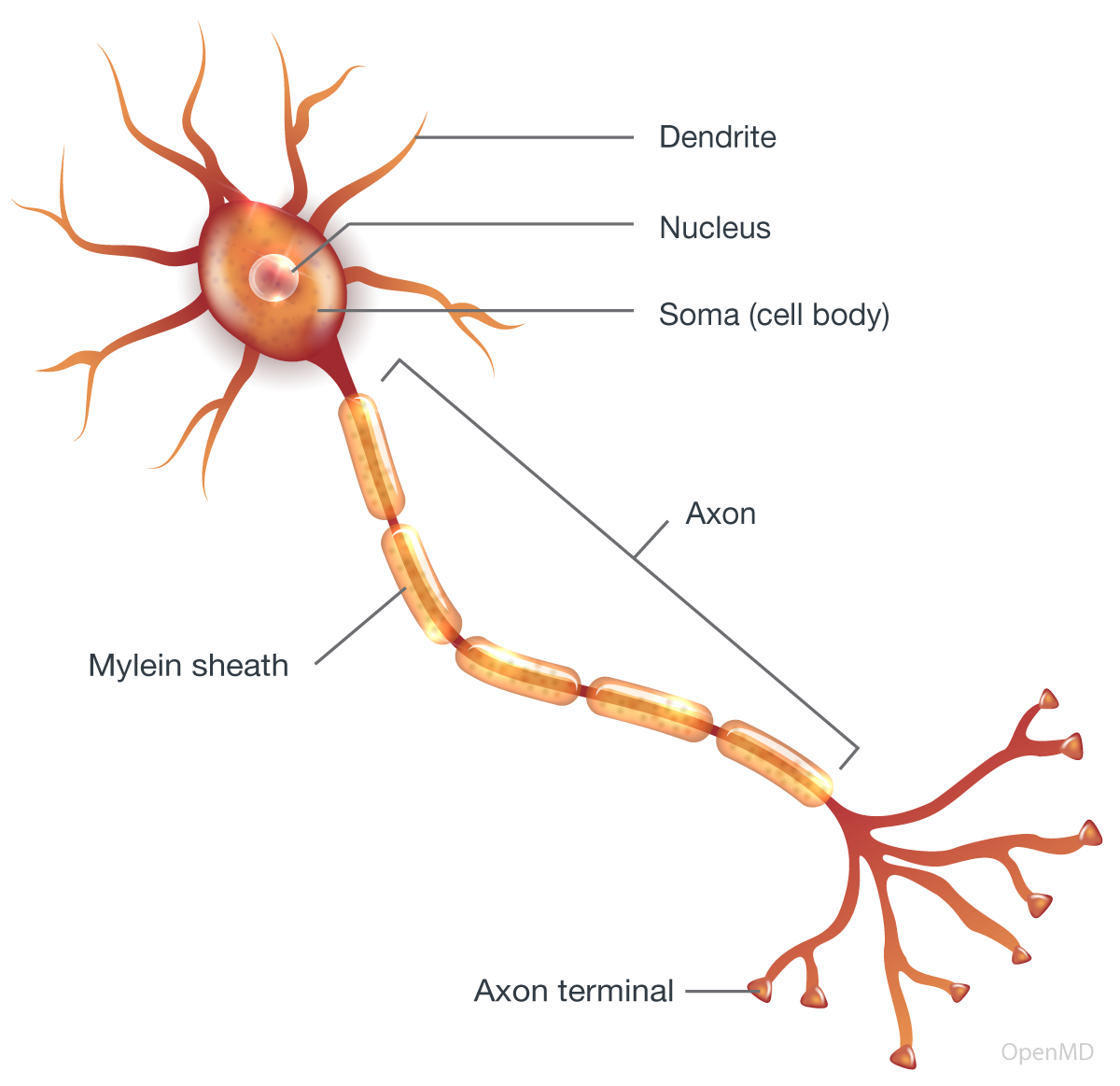
Multipolar neuron with myelin sheath
Neurons
Neurons (nerve cells) are the main functional unit of the nervous system that generate and propagate electrical signals (called action potentials) along their axons to communicate with other cells. They have specialized structures that include the soma (cell body), dendrites, the axon (a long cable-like projection), and axon terminals.
- Soma: The soma is the cell body that contains the nucleus, which houses the genetic material of the cell and other organelles (tiny structures within cells that perform specific functions).
- Dendrites: Branch-like structures that extend out from the cell body, dendrites receive signals from other neurons, acting as the main input sites for the cell.
- Axon: A long, thin, cable-like extension from the cell body, the axon carries electrical signals to the axon terminals.
- Axon terminals: The button-shaped end points of the axon, axon terminals release neurotransmitters (brain chemical messengers), which are received by other neurons or muscle fibers.
Glial Cells
Glial cells provide support and protection for neurons. They include astrocytes, oligodendrocytes, Schwann cells, microglia, and ependymal cells.
- Astrocytes provide structural support and help maintain the chemical environment around neurons.
- Oligodendrocytes form the myelin sheath around axons, providing insulation that allows for faster and more efficient transmission of action potentials.
- Schwann cells form the myelin sheath around axons in the peripheral nervous system.
- Microglia protect the brain and spinal cord from injury and infection.
- Ependymal cells line the cerebral ventricles (fluid-filled spaces within the brain) and the spinal cord’s central canal. They produce cerebrospinal fluid (CSF), which cushions, nourishes, and protects the brain and spinal cord.
Physiology
Signal Transmission and Modulation
Neurons transmit information by sending electrical impulses (action potentials) along their axons to synapses, tiny gaps between neurons where neurotransmitters are released and received by the dendrites or cell bodies of neighboring neurons. This process continues until the signal reaches its intended target, such as a muscle or gland.
The effect of a neurotransmitter depends on the type of receptor protein on the postsynaptic neuron. Receptor proteins can be either ionotropic or metabotropic.
- Ionotropic receptors are channels that open or close when a neurotransmitter binds to them, changing the postsynaptic neuron’s membrane potential (the electrical balance across cell walls, influencing the readiness of neurons to transmit signals in the brain and body).
- Metabotropic receptors are complexes that trigger metabolic changes in the postsynaptic neuron when a neurotransmitter binds to them, involving a second messenger molecule.
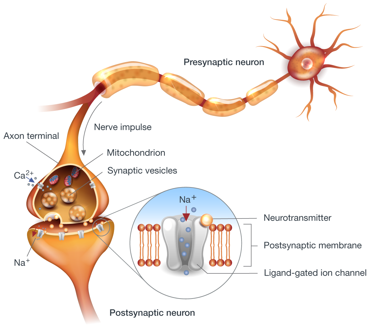
Different neurotransmitters and receptors can have excitatory or inhibitory effects on the postsynaptic neuron, modulating (changing) its activity.
Neuroplasticity and Growth
Neuroplasticity refers to the brain’s ability to modify its structure and function. This plasticity operates differently in brain development vs. adulthood.
- In development, neuroplasticity enables dramatic changes, such as neurogenesis (the birth of new neurons), neuronal migration, axon/dendrite growth, synapse formation, and large-scale changes in brain organization and acquisition of functions like vision and language.
- In adulthood, neuroplasticity is more limited and mainly involves subtle changes in synaptic strength, although there is evidence that neurogenesis can occur in parts of the adult brain. Attempts at large-scale axon regeneration have been challenging.
- Myelination of axons (growth and repair of the myelin sheath) appears plastic in both development and adulthood, particularly in the peripheral nervous system.
Neuroplasticity can be adaptive, but also maladaptive, contributing to some neurological and psychiatric disorders.
From the flutter of a heartbeat to the spark of an idea, the human nervous system is the master controller. This intricate network of nerves not only regulates our vital functions but also fuels our senses, thoughts, and actions. It is our bridge to the world, allowing us to perceive, respond, and shape our reality.
Additional resources:
- Introduction to the Nervous System by the National Cancer Institute
- Structure and Function of the Nervous System by OpenStax
References:
- Innocenti GM. Chapter 1 - Defining neuroplasticity. Handbook of Clinical Neurology. ScienceDirect. January 1, 2022.
Contributors:
- Ari Magill, M.D.
Editors:
- Brian Sullivan, MD