Human Skeletal System
The skeletal system provides the structural framework for the human body. It consists of bones, joints, cartilage, ligaments, and tendons, all of which work together to provide support, protection, and movement. The skeletal system is closely integrated with the muscular system.
The functions of the skeletal system include the following:
- Support: Provides a framework that supports the body and maintains its shape.
- Protection: The skull, spinal column, rib cage, and pelvic girdle enclose and protect internal organs.
- Movement: In conjunction with the muscular system, the skeletal system enables movement at the joints.
- Hematopoiesis: Some bones contain marrow, which produces blood cells.
- Mineral and Fat Storage: Bones store minerals, such as calcium and phosphorus, important for bone health and bodily functions. Fat is stored in yellow bone marrow.
Bones
Bones, comprising a complex structure of mineralized connective tissue, primarily consist of calcium and phosphorus in a matrix of collagen fibers. They provide the strength and flexibility that are essential for supporting the body’s structure and facilitating movement. There are several shapes of bones in the human body:
- Long bones: Characterized by their length and primarily responsible for weight bearing and movement. Examples include the femur, tibia, and humerus.
- Short bones: Compact bones that provide stability and support, such as the bones of the wrist and ankle.
- Flat bones: Provide protection and support for the body’s organs. Examples include the scapula and the bones of the skull.
- Irregular bones: These have complex shapes and serve various functions. Examples include the vertebrae and the bones of the face.
- Sesamoid bones: Small bones, such as the patella (knee cap), embedded within muscles or tendons. The number of sesamoid bones varies from person to person.
There are two primary types of bone tissue: compact and spongy bone. Compact bone, or cortical bone, forms the exterior of most bones, providing strength. It consists of densely packed layers composed of osteons, containing mineralized matrix and collagen. Compact bone encircles central canals that contain blood vessels and nerves.
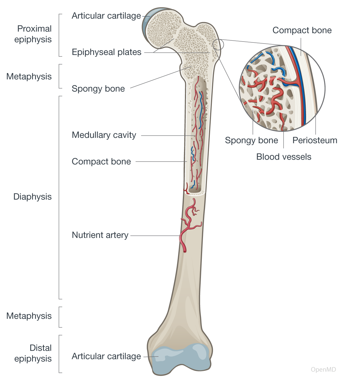
Spongy bone, or cancellous bone, is found in the epiphyses of long bones, within flat bones, and in vertebral bodies. It is lighter and more porous than compact bone, with a trabecular network that provides support and reduces weight. These trabeculae, surrounded by marrow (red or yellow), aid in blood cell production in red marrow and fat storage in yellow marrow.
Joints
Joints are the connections where two or more bones meet, allowing for movement. Based on their mobility and structural characteristics, there are three main types of joints: fibrous (synarthrodial), cartilaginous (amphiarthrodial), and synovial (diarthrodial).
- Fibrous Joints (Synarthrodial): Immovable (or nearly immovable) joints connected by fibrous tissue, such as the sutures between the cranial bones and the syndesmoses between the tibia and fibula.
- Cartilaginous Joints (Amphiarthrodial): Slightly movable joints connected by cartilage, such as the joints between the vertebrae in the spine.
- Synovial Joints (Diarthrodial): Characterized by a fluid-filled joint cavity that allows for a wide range of movements. Synovial joints can be further classified by their shape and permitted range of motion. This classification includes:
- Ball-and-Socket Joints (Spheroid): Allow for a wide range of movement in almost all directions (rotation, abduction, adduction, flexion, and extension). Examples include the shoulder and hip.
- Condyloid Joints: Allow for flexion, extension, abduction, adduction, and circumduction. These joints permit movement but not rotation. Examples include the wrist, knuckles, and toes.
- Hinge Joints (Ginglymus): The elbow and knee are examples of hinge joints, which enable movement similar to the opening and closing of a hinged door (primarily flexion and extension).
- Pivot Joints (Trochoid): Allow for rotational movement around a single axis. A prime example is the joint between the first and second cervical vertebrae (the atlas and axis) in the neck that allows the head to turn side-to-side.
- Saddle Joints: Allow for movements along two perpendicular axes (flexion, extension, abduction, and adduction). Examples are found in the thumb, thorax, and heel.
- Plane Joints: Allow for gliding movements where the bone surfaces slide past each other. The joints between the carpal bones in the wrist are plane joints.
Cartilage
Cartilage is a type of connective tissue that provides cushioning and reduces friction between bones. It covers the ends of bones within the joints and is essential for smooth and pain-free movement. There are three main types of cartilage: hyaline cartilage, fibrocartilage, and elastic cartilage.
- Hyaline cartilage: The most common type of cartilage, it is found in the knees, hips, rib-to-sternum connections, nasal cartilage, and joint surfaces. It enables smooth joint movement.
- Fibrocartilage: Denser, stronger cartilage in load-bearing areas such as the spinal discs, knee menisci, and pubic symphysis. It absorbs shock and resists compression.
- Elastic cartilage: Flexible, strong cartilage, found in the external ear, epiglottis, and larynx, that maintains shape while allowing flexibility for function.
Ligaments and Tendons
Both ligaments and tendons play important roles in maintaining the stability and integrity of the skeleton. Ligaments, strong fibrous bands of connective tissue, primarily connect bones to bones and stabilize joints. This stability prevents joint dislocations and enables finer motion control. Examples of ligaments include the cruciate ligaments in the knee, which control movement, as well as the collateral ligaments, which provide lateral stability. Tendons are similar in structure to ligaments, but they connect bones to muscles and transmit the force generated by muscle contraction to the skeleton.
Human Skeleton
The skeleton is divided into two main regions: the axial skeleton and the appendicular skeleton. The axial skeleton forms the central axis of the body and protects the critical organs. The appendicular skeleton includes the bones that are appended to the axial skeleton. These appendages enable the body to move and interact with the environment.
Axial Skeleton
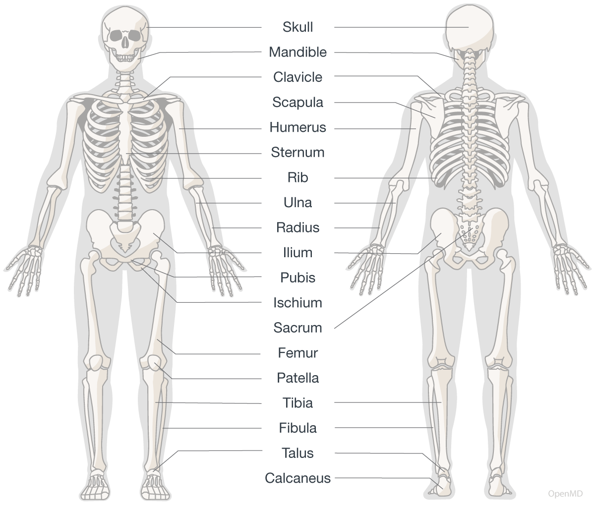
The axial skeleton is composed of 80 bones and forms the central axis of the body. It consists of the skull, vertebral column, rib cage, and sternum.
- Skull: The skull is composed of the cranium, facial bones, and hyoid bone.
- Vertebral column: Consists of 24 separate vertebrae, divided into 5 regions: cervical, thoracic, lumbar, sacral, and coccygeal.
- Rib cage: Formed by 12 pairs of ribs and their associated costal cartilages.
- Sternum: Flat bone located at the center of the rib cage.
Appendicular Skeleton
The appendicular skeleton consists of 126 bones that form the upper and lower limbs as well as the pectoral and pelvic girdles.
- Upper limbs: Composed of the humerus, ulna, radius, carpals, metacarpals, and phalanges.
- Lower limbs: Composed of the femur, tibia, fibula, tarsals, metatarsals, and phalanges.
- Pectoral girdle: Composed of two clavicles and two scapulae.
- Pelvic girdle: Composed of two coxal bones that articulate with each other anteriorly at the pubic symphysis.
Development and Growth
The skeleton undergoes several stages of growth and development, including ossification (the process of bone formation), elongation, and remodeling. Hormonal and genetic factors play a crucial role in the growth and development of the skeleton, and environmental factors, such as diet and exercise, can also influence its growth and health.
In childhood, red bone marrow predominates. It actively produces blood cells, including red and white blood cells, and platelets. As individuals mature, some red marrow is converted to yellow marrow, which is primarily fatty and does not produce blood cells. Yellow bone marrow can be replaced by red bone marrow when there is an increase in the body’s need for blood cells. This process is known as reconversion.
During childhood, the rapid growth and elongation of bones are facilitated by specialized structures known as growth plates, located at the ends of long bones. As the child grows, the bone lengthens at the growth plate. At maturity, the cartilage in the growth plates is gradually replaced by bone, and the plates eventually fuse, which stops further lengthening of the bone.
The skeleton is constantly undergoing maintenance and repair through a process known as remodeling. This involves the removal of damaged or worn bone tissue by specialized cells called osteoclasts, followed by the formation of new bone tissue by cells called osteoblasts. Osteocytes, mature bone cells within the matrix, are crucial in remodeling. They detect stress on bones and signal osteoblasts and osteoclasts to start remodeling as needed. Bone remodeling is a complex process that is regulated by a variety of hormonal factors, including parathyroid hormone and calcitonin. It is influenced by nutritional intake, including calcium and vitamin D, and mechanical stress from physical activity. This dynamic process balances bone formation and resorption, which is crucial for maintaining bone strength and health.
Additional resources:
- Introduction to the Skeletal System by the National Cancer Institute
- Human Skeletal System by Britannica
Contributors:
- Teresa Roberts, DNP
Reviewer:
- Brian Sullivan, MD